Cambridge Colour Testing Method
A Review on the Cambridge Color Testing Method
 To evaluate a person's color vision and discrimination is an essential part of a routine vision and eye exam. With the increasing prevalence of hereditary color vision deficiency, color vision disorders can be diagnosed by an ophthalmologist using a variety of available tests.
This article provides an overview of the structure and function of a selection of color vision tests that have been performed manually and partially modified into a computerized version.
In various activities, as well as in congenital or acquired color vision deficiency (CVD), color vision tests and examinations are used not only for the early detection of CVD, but also for monitoring the progression or remission of the disease. Loss of color vision can sometimes occur due to the side effects of certain medications used to treat diseases such as diabetes, high blood pressure, HIV and AIDS, among others.
This test can be assessed qualitatively and quantitatively through tests and grouped them together as arrangement tests, matching tests, vocational tests or pseudoisochromatic plates. Simple or more sophisticated computerized methods such as the Cambridge Color Test (CCT) are sometimes used to assess color function or discriminate.
Literature review
The human retina comprises two classes of photoreceptors: rods and cones. Rods are responsible for vision in dim light, and cones mediate vision in bright light to enable the perception of color. Individuals with normal color vision have three types of cone photoreceptors: short wave sensitive or blue, middle wave sensitive or green, and long wave sensitive or red cones.
All such individuals have normal trichromatic color vision. Individuals with severe color vision defects usually either have non-functional red (protanopes) or green (deuteranopes) cone photoreceptors and have dichromatic rather than trichromatic color vision. Those with milder color vision defects usually have either their red or green cone photoreceptor pigments replaced by an anomalous pigment with altered spectral sensitivity; such individuals are classified as having protanomalous or deuteranomalous trichromatic color vision.
One of the main applications for color vision screening is to assess and detect deficiencies that occur in the visual system as a result of congenital or pathological causes. The evaluation of trichromatic color vision in individuals is based largely on color discrimination and color matching. As human retinae include three types of wavelength sensitive cones, color is a trichromatic process and various tests have been developed to assess trichromatic color vision by means of arrangement, matching, and vocational and pseudo-isochromatic plates.
Normal trichromatic vision can be defined as the ability to distinguish and discriminate light on the basis of wavelength composition; that is, independent of its intensity. Color perception is dependent on the absorption of light by three classes of photoreceptor cones in the retina, namely short (S-), medium (M-) and long (L-) wavelength sensitive cones, whose peak sensitivities are ≈420 nm, ≈ 530nm and ≈560nm respectively.
Each cone of the above classes obeys the Principle of Univariance; that is, the absorption of a long wavelength (low frequency, low energy) quantum has the same effect on a receptor as the absorption of a short wavelength (high frequency, high energy) quantum. It is the probability of absorption that changes photoreceptor sensitivity. Any single photoreceptor is 'color blind', since an appropriate combination of wavelength and radiance can produce an identical neural response.
No individual receptor can differentiate between changes in wavelength and radiance and, although the probability of that cone being stimulated by a photon of light depends on wavelength and radiance, the output can only vary by the degree of depolarization within the receptor and is independent of wavelength .
Using this principle, a comparison of signals from two or more cone classes (each with a different spectral sensitivity) is required to make a differentiation between wavelength and radiance.
All possible colors can be plotted in a three-dimensional (3D) color space because the normal retina contains only three classes of cones, each obeying the Principle of Univariance. The three axes of a 3D graph or stereo-pair can be used to represent different quantities relating to color vectors.
For example, a color vector could be plotted in terms of red (R) or L-cone input, green (G) or M-cone input and blue (B) or S-cone input. The color space is then described as RGB-space or LMS-space. A given color vector [eg cw = (1 1 1)- or white (subscript, w)] can be plotted as a point in this 3D space and, as radiance is changed, the point (and its color) will move along a vector that extends from the origin [usually but not always the color black with 1 × 3 color vector ck = (0 0 0)-, which is essentially an absence of color].
The angle of the color vector relative to the axes of the color space corresponds to its chromaticity, where chromaticity can be defined as the quality of a color or light with reference to its purity and its dominant wavelength.
Munsell, Ostwald and the Commission Internationale de l'Éclairage (CIE) describe color space with different parameters,14 and the CIE (1976) system which is used with the CCT, uses a parameter Y to measure brightness and parameters such as x and y (or u and v or u' and v') to specify the chromaticity.
Mollon (2000) explains that dichromacy of color discrimination is the basic principle underlying the CCT. A pair of physical stimuli can be chosen that yield the same chromaticity in two of the three classes of cones, but differ with regard to the remaining class. In alternating such stimuli, one can establish the integrity of the one isolated class of cones.
For instance, consider a point that lies in the plane of the chromaticity of the medium wavelength and long wavelength cones. If a line were to pass orthogonally through that point, lights along that line would vary only in the line of the chromaticity of the short wavelength cones. This is called the 'tritanope confusion line', as someone who lacks short wavelength cones will confuse chromaticities in that line or direction. A tritanope, deuteranope or protanope is a person who confuses chromaticities along the short, medium or long wavelength confusion lines respectively.
Cambridge Color Test
The CCT is an example of a computerized test. Computerized color vision testing offers the advantage of being able to adjust the difficulty depending on the patient's performance, as well as randomizing plates to avoid the patient from recalling previously previewed plates.
The design of the CCT uses a computer version of pseudo-isochromatic plates and combines the Principles of Chibret and Stilling. Chibret's Principle (1877) states the possibility of varying the chromatic difference of the target and field dynamically and adaptively, along different directions in color space. Stilling (1877) achieved non-hue noise by varying the size and luminance of the target and background elements.
Each of the computer pseudo-isochromatic plates in the CCT contains a Landolt C; that is, each stimulus is a large colored letter C of specific hue and luminance that is presented in four different orientations; the opening in the letter can be up, down, right or left, which is embedded in a background comprising circles of varying size, color and luminance.
The CCT stimuli backgrounds eliminate luminance or contour cues, and the figures (the test stimuli of Landolt letters) must be identified solely by their hues. As mentioned, this is accompanied by non-hue noise achieved by varying the luminance and size of compositional elements in accordance with Stilling's Principle.
The CCT controls the color spaces (using the CIE u'v' [1976] chromaticity diagram) of the figures or stimuli (the Landolt letters) and the background and presents a random presentation of figure-background chromaticity differences. The patient presses one of four keys of a hand-held input device to indicate the openings of successive stimuli (the Landolt C) presented, making this task cognitively simple.
Estimation of discrimination thresholds uses a staircase procedure which is an integral part of the two CCT tests, namely Trivector and Ellipses. The Trivector test makes use of the protan, deutan and tritan confusion lines to probe the sensitivity of the long, medium and short wavelength cones.
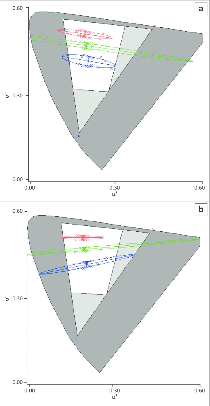
Each target differs from the background along one of the three lines in color space; that is, of either the deutan, protan or tritan confusion lines. The Ellipses test determines the parameters of the three MacAdam ellipses that lie along the same tritan confusion line. Discrimination ellipses show a loss of sensitivity for a range of directions around the background chromaticity, with the long axis of the ellipse being indicative of the type of loss.
The three MacAdam ellipses that are measured in the Ellipses test correspond to three different background chromaticities, and the ellipses are represented graphically using best-fit closed curves to the thresholds (indicated with crosses) measured for different directions relative to the background color concerned. Generally, protanopes display ellipses with a bottom-left slant and deuteranopes develop a top-left slant.

Normative data for normal trichromatic color vision patients using the CCT were published by two groups of researchers from Brazil. One group from Sao Paulo used the CCT v.2.0 with a VSG S card and Sony FD Trinitron color monitor; the second group from Belem used a self-built system for the IBM RISC 6000 workstation and IBM 6091 19i color monitor. The results from both studies for the Trivector test were similar, and the absence of significant statistical difference between the two sets of data indicates the reliability of results produced by the CCT.
The CCT has been used in a wide variety of studies; one example is the determination of color discrimination losses in patients with chronic diseases such as multiple sclerosis. In such cases, the CCT helped in characterizing vision losses in both red-green and blue-yellow discrimination, indicating that both parvocellular and koniocellular visual pathways were affected.
The CCT also identified the relationship between these losses and the degree of optic nerve involvement. Parvocellular and koniocellular cells are located in the lateral geniculate nucleus (within the thalamus of the brain) which is the primary relay center for visual information received from the retina of the eye. Parvocellular cells essentially respond to long and medium wavelength ('red' and 'green') cones whilst koniocellular cells are short wavelength 'blue' cones; the three types of cones are necessary for the perception of color and form; that is, including gross and fine detail.
A color vision test such as the Ishihara is not designed for detecting defects along the blue - yellow axis. However, the CCT has been developed to overcome difficulties in isolating the chromatic channels of the visual system, thus making it a versatile tool in both research and detection of a multitude of color defects across all chromatic channels such as in multiple sclerosis.
The CCT was also adapted to be used on animals such as squirrel monkeys. Animal color vision testing occurs to determine the presence, depth and acuteness of color vision as well as to assess differences within species. Unlike other color vision tests, this computer-based test allows researchers to modify the test so that the CCT can easily be used to evaluate color vision in different species.
The adapted version of the CCT is more efficient than other more conventional methods because it eliminates the need to equate the luminance of each chromaticity tested. Results proved reliable because the stimulus parameters could be adjusted so that the animals were not able to use luminance differences to make correct discriminations.
In Mancuso et al., two squirrel monkeys were used and different hues were tested. Three separate color vision tests were completed by each of the two monkeys and thresholds obtained were plotted on CIE x, y graphs where the x-axis showed the 16 different hues specified in terms of dominant wavelength of the most saturated color tested, and the y -axis showed the corresponding thresholds for each hue.
The time required to test 16 hues took about 2–4 months; therefore, the results represented color vision testing conducted over a year. Their graphs identified similar results for each test, and repeated measurements were highly consistent, showing that results produced by the CCT are highly repeatable even though these results were obtained from monkeys.
In a study in 160 normal trichromats by Paramei, using the CCT to assess color discrimination across four life decades, the results indicated no significant differences in the CCT outcomes between two groups of normal trichromatic young adults (20–29- or 30–39- year-olds). There was, however, individual variability within each group, which increased with age. Also, for normal trichromats of the same age range, binocular summation, eye dominance and learning do not have an effect on color discrimination thresholds as determined by the CCT.
Computer-based color vision tests Computer-based color vision tests
In addition to the CCT, another computer-based approach for screening color deficiencies was done by presenting the Ishihara plate on a CRT monitor. Technical restrictions of CRTs in computer monitors imply that not all perceivable colors can be adequately presented on a monitor.
The generation of color is done by mixing light emitted by three phosphors in the red (R), green (G) and blue (B) regions; this technique leads to an RGB coordinate system to describe colors on a computer monitor by the intensities of the cathode rays emitting from the three phosphors.
Certain shades of orange, yellow and blue-green colors cannot be represented on a CRT monitor, which leads to the assumption that the spectral emission of Ishihara plates on a CRT monitor will be different from the spectral emission of reflected daylight on paper plates. This effect is caused by the many orange and red dots on the Ishihara plates. An identical spectral emission of the reflected daylight of the plates and the emission from a CRT monitor is not possible.
Nevertheless, calibration of the monitor allows a controlled spectral emission for the Ishihara plates represented on a CRT monitor in contrast to uncontrollable daylight. Results of tests done with both the Ishihara plates and the PC-based test were both comparable and reliable for screening purposes. The fact that a monitor can be controlled allows a higher degree of repeatability of the examination results using emitted light by controlled constraints.
This control can be seen as an advantage of a CRT monitor presenting the Ishihara plates, because daylight cannot be controlled.
Other technologies besides CRT can be used in monitors, for example, light emitting diodes (LED) and LCD; computer color tests have been designed for such monitors but are not addressed any further in this review.
